From Friday, April 19th (11:00 PM CDT) through Saturday, April 20th (2:00 PM CDT), 2024, ni.com will undergo system upgrades that may result in temporary service interruption.
We appreciate your patience as we improve our online experience.
From Friday, April 19th (11:00 PM CDT) through Saturday, April 20th (2:00 PM CDT), 2024, ni.com will undergo system upgrades that may result in temporary service interruption.
We appreciate your patience as we improve our online experience.
Please complete the following information
University and Department: School of Life Sciences and Biotechnology, Shanghai Jiao Tong University
Team Members: Xiaowei Wu, Tobias Braeuler, Cuixia Dai, Peng Xi, Chuanqing Zhou, Qiushi Ren
Faculty Advisors: Peng Xi
Primary Email Address: xipeng@sjtu.edu.cn
Primary Telephone Number (include area and country code): +86 21 3420 4076
Project Title: Frequency-Domain Optical Coherence Tomography (FD-OCT)
List all parts (hardware, software, etc.) you used to design and complete your project:
NI DAQ PCI 6731
NI IMAQ PCI-1428 image acquisition card
LabVIEW 8.5
Describe the challenge your project is trying to solve.
OCT is a high-resolution optical cross-sectional imaging technology. We need to develop a FD-OCT system for the imaging of the retina of the human eye.
In our experiment, the following challenges need to be solved:
(1) a constant timing clock for each galvanometric scanning.
(2) Two voltage signals to drive the galvanometer pair and continuous galvanometric scanning driving;
(3) Synchronization with the line-CCD camera;
(4) Post-processing of the interferogram for visualization;
(5) A user-friendly GUI for easy operation of the system.
Describe how you addressed the challenge through your project.
In our system, a FD-OCT is constructed for the imaging of the retina of the human eye. (Fig.1)
A broad band light sources beam is employed as the low coherent light source. The incidence was split in a fiber coupler and focused on the background of the eye and on a reference mirror, respectively. The reflected beams are recombined in the fiber coupler and interfere there, coding the information of the retina in the interferogram. Afterwards, the interferogram is recorded by a grating-based spectrometer. The spectrometric data was taken with a line CCD camera (Aviiva e2V). Together with NI IMAQ PCI-1428 image acquisition card, the data for each A-scan is recorded and then processed for visualization by a custom-designed program written with Labview.
Each shot gives us on depth scan (A-scan) of the eye, with two galvanometric scanning mirrors the beam is scanned across the surface of the retina to record A-scans at different positions to reconstruct the 3-D morphological structure of the retina in vivo (Fig. 2).
At several core parts of our system, NI products are used:
l First of all, the scanning mirror in the galvanometer are driven and controlled by a NI PCI 6731 data acquisition card, allowing an easy to control and precise beam positioning.
l The intferogram, recorded by a line-scan CCD camera, is transferred to the PC by use of the NI IMAQ PCI 1428 image acquisition card, allowing an exact triggering and fast image transfer.
l The mirror in the reference arm is mounted on a motorized actuator stage, making fine adjustment of the mirror position possible. The stages motion controller is connected to a NI PCI-GBIP card.
All of these parts are controlled by NI Labview 8.5. The post processing is also done with Labview. Furthermore, we also designed a Labview based, custom made, graphical user interface, allowing an easy access to the system and suiting the demands of medical professionals. (Fig.3)
Please upload photos below:
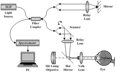
Fig. 1 Diagram of the FD_OCT system.
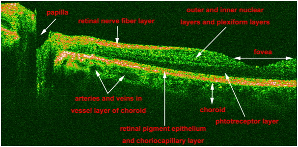
Fig. 2 Our OCT imaging result revealed the biological structures of the human retina.
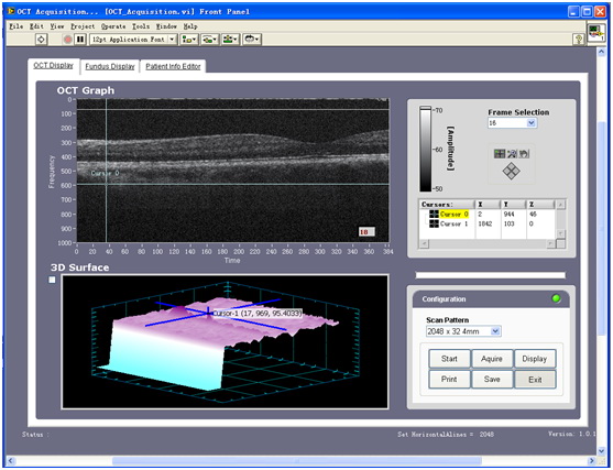
Fig. 3 The FD-OCT GUI.
Please insert video below:
[Click on video icon on task bar]
\\spanky\CDG-Shared\China\Web\video.avi
or
